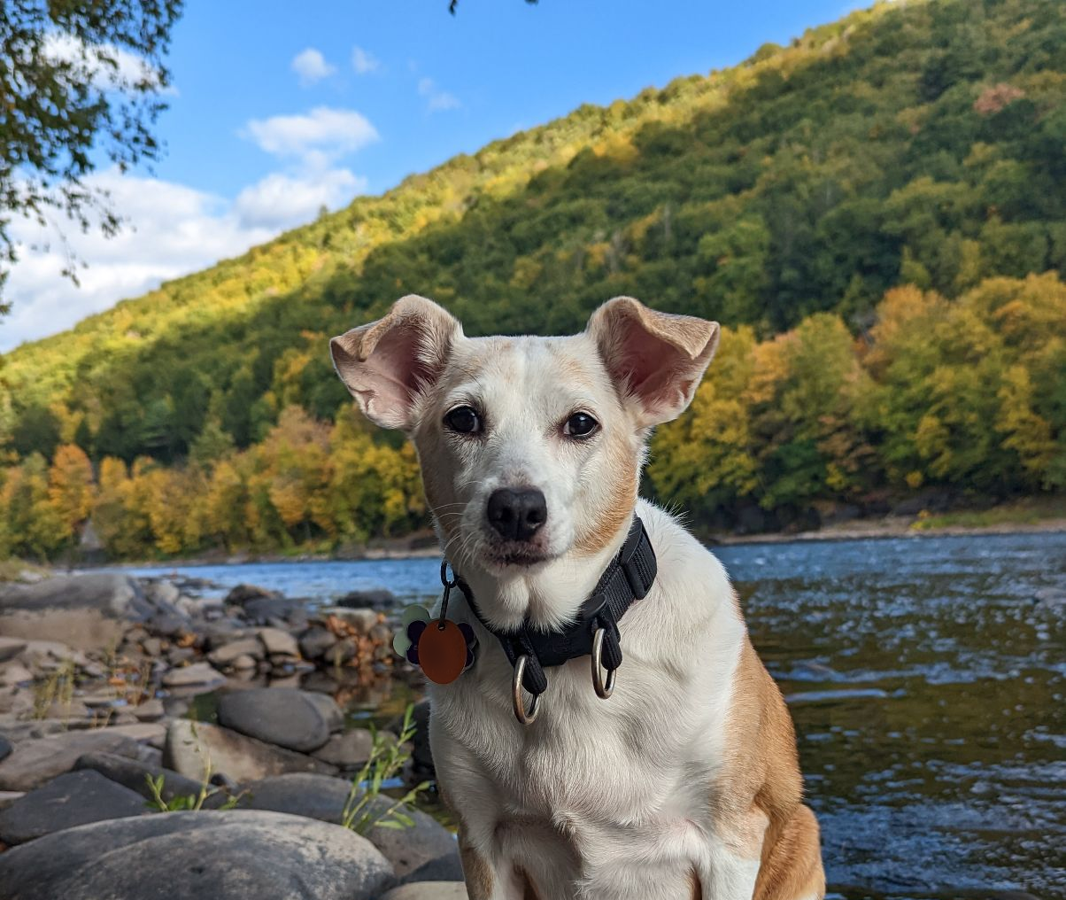
Kimmy, a 14 year old Terrier mix, was in deep trouble. She had seen multiple vets at several practices. Long story short, after an ultrasound and a cat scan, she had been diagnosed with a large cancerous tumor of the liver.
Her signs appeared out of the blue. Instead of being her usual energetic and bubbly self, she wouldn’t walk or move, she peed on the floor, and “her belly moved oddly” recalls her owner.
After a visit to the ER, it was determined that she had “free fluid” and an incredibly large mass in her belly.
The discharge papers said she could face “possible instant death.”
“We were devastated,” commented her owner.
And that’s how we met, after the owners “researched so many surgeons in PA, NJ, and NY.”
We had a long conversation about the risks of surgery. The owners asked multiple, excellent questions about surgery and anesthesia. I’ve rarely been quizzed so diligently by a pet owner!
PLEASE NOTE that the upcoming picture is very graphic !!!
“Leading up to surgery, I tried to treasure every moment, cuddling her more often. I took lots of photos and videos.”
The day of surgery, after getting perked up with IV fluids, Kimmy was placed under anesthesia.
We opened her belly up, and sure enough, we found a huge, very firm mass in the liver.

Despite the ultrasound and the CAT scan findings, we realized that there was no way to remove the mass safely.
It was attached to multiple organs and vital structures.
Among others, the mass was attached to a large artery, called the aorta, and a large vein, the vena cava.
It was firm, apparently with some “solid” areas and others that felt full of fluid.
As promised before surgery, we called the owners in the middle of the surgery to discuss our findings.
The situation was not good. Together, since removing the mass was not possible, we decided to at least take some biopsies so we knew what we were dealing with. That would respect the owners’ main wish: to get Kimmy back home – no matter what.
Kimmy’s owner recalls: “When you said the mass was really big and too risky to take out due to location, I felt like the bad news I’d been bracing for was becoming reality. I wanted a solution and deeply wanted some hope or sign that this wasn’t going to be it for her, that she had several more months to live.”
In addition, “Things seemed to be going downhill faster than we were ready for, and it was terrifying. At the end of the call, knowing the mass would still be there made me feel like we had lost some options, and options mean more chances of survival.”
So we took a few biopsies of the evil mass. The last biopsy site revealed that a large part of the mass was full of yucky, brown fluid.
We removed a surprising 450 ml of fluid, which is the same as 15 fluid ounces, or almost 2 cups!
Keep in mind that Kimmy weighed 24 pounds (11 kg).
As we removed the fluid, we saw the mass literally shrink to a fraction of its original size!
Before we could even stitch everything back up, the owner called the clinic to ask if we could euthanize Kimmy before she woke up.
Can you imagine the emotional rollercoaster?
I asked the nurse to relay a simple message: “Please hang in there, I absolutely do not recommend euthanasia at this point.”
As soon as Kimmy woke up, I called the owners to explain our findings. It still could be cancer, but we should at least provide her with some temporary relief.
After all, this huge mass was causing a lot of pressure and must have caused significant pain.
“When you called us back after surgery, you gave us hope,” said the owner.
After one night in the hospital on IV fluids, antibiotics and pain medications, Kimmy went home to recover.
About a week after surgery, the biopsy report came back. What do you think it said?
Despite all of the previous tests, despite what everybody said before surgery, the mass was not cancer.
It was benign !!!
Of course, I didn’t believe the pathologist, so I called him.
He explained that everywhere he looked, there was no sign of cancer in our biopsies!
The tentative diagnosis is an unusually large “biliary cyst,” in other words, a cavity full of bile.
In addition, remember the brown fluid looked “yucky” (a highly scientific description)?
The pathologist thought was based on chemical analysis of the fluid, the bile had been sitting there for a while.
At the end of the call, we agreed that this cyst probably grew slowly over time.
Initially, it was small. Then it grew bigger and bigger, until Kimmy couldn’t take the pain anymore.
We then agreed that a possible long-term solution could be to drain the cyst through the skin, under the guidance of an ultrasound, if Kimmy has similar signs in the future.
I called the owner to share the great news.
She remembers: “Whew! I was expecting bad news, and received the best news of the year. The mass was benign, I was so incredibly relieved!!! The rollercoaster was so crazy and it seemed like the nightmare was finally over.”

At home, “Kimmy was doing so well! She was back to her old self already. I am positively over the moon that Kimmy will be with us for a long time after this wild storm. We have many more adventures to get to together.”
And 6 months after surgery, Kimmy is reportedly still doing great…
If you would like to learn how we can help your pet with safe surgery and anesthesia, please contact us through www.LRVSS.com
Never miss a blog by subscribing here: www.LRVSS.com/blog
Phil Zeltzman, DVM, DACVS, CVJ, Fear Free Certified
Pete Baia, DVM, MS, DACVS
