Brodi, a 14 year old miniature poodle, had a big mass on his chin. In fact, he had 2 masses, right next to each other.
FAIR WARNING.
The pictures below can be considered graphic by some sensitive readers, so please don’t look if you think you might feel queasy!!!
A smaller mass had been removed a while back, but it slowly grew back.
It had been diagnosed as a basal cell tumor, a common tumor that can be benign or malignant.
In Brodi’s case, it was fortunately benign but “locally invasive.”
It became larger and larger, to the point that it was bleeding all the time.
The owner questioned removing the mass in a 12 year old dog.
But Brodi lived to be 13, and the mass grew bigger.
So his owner questioned removing it in a 13 year old dog.
But Brodi lived to be 14, and the mass grew bigger.
So his owner questioned removing it in a 14 year old dog.
What helped her make the decision is that Brodi’s preop blood work was very good for a 14 year old.
After a lot of soul-searching, she decided to consider surgery and we met for a consultation.
Not surprisingly, I said I would be willing to tackle it.
Anesthesia is very safe these days, and Brodi was overall in good health.
As for surgery, it was clearly a challenge, but after researching various option, I came up with a reasonable plan of action.
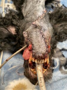
Below are a few pictures, from various angles, to give you an idea of the masses in 3D.
AGAIN, YOU HAVE BEEN WARNED, THESE PICTURES ARE NOT FOR THE FAINT OF HEART.
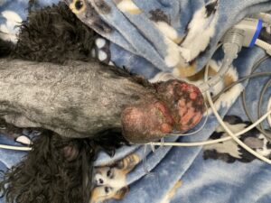
View from the top showing the “double mass”
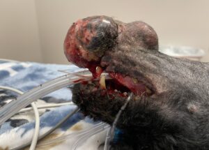
View from the right side showing the “double mass”
View from the left side showing the “double mass”
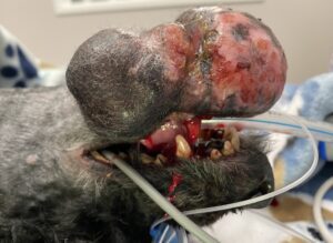
Once the masses were removed, there was physically no way to close the skin.
The plan was to “rob (skin) from Peter to give to Paul.”
Before we explain how we solved that problem, let’s go over 2 quick definitions:
. a skin graft is taken from one body part, lifted off entirely, and applied elsewhere.
. a skin flap is taken from an area close to where it’s needed, turned around a specific amount, and stitched in the defect.
In Brodi’s case, we would use a skin flap by borrowing skin from the neck, and use it to cover the big gap under his lower jaw.
The key was to find enough skin to avoid any pressure along any of the incisions.
One of the risks was that if Brodi stretched his neck, he could rip some or all of the stitches.
In the end, the masses came out nicely, and 100% of the skin defect was closed.
This is how Brodi looked at the end of surgery, again from various angles.
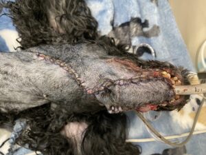
View from the top after the skin flap was sutured
View from the front after the skin flap was sutured
And this is how Brodi looked after 3 weeks of healing and TLC at home.
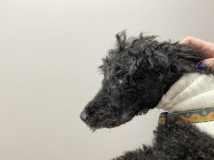
His owner concludes: “Things are so much better now. Brodi looks great. I know I did the right thing!”
If you would like to learn how we can help your pet with safe surgery and anesthesia, please contact us through www.LRVSS.com
Phil Zeltzman, DVM, DACVS, CVJ, Fear Free Certified